facs buffer flow cytometry
This Flow Cytometry Staining. Built on a foundation of excellence experience and expertise the BD FACSLyric is a new standard for.

Single Cell Rna Expression Analysis Using Flow Cytometry Based On Specific Probe Ligation And Rolling Circle Amplification Acs Sensors
Ad Run sticky samples at high flow rates with a system that is exceptionally clog resistant.

. Stop cell lysis by adding 10ml Cell Staining Buffer to the tube. Ad Chemical Biochemical Products Kits Used in Scientific Research. Place samples in 12 x 75 mm Falcon tubes and analyze by flow cytometry as soon as possible within 1 hour.
If titrating antibodies and storing aliquots of the same add sodium azide in the storage buffer at 009. Compatible with optional autosamplers and robotic integration for walk-away automation. Resuspend cells with 052 mL FACS buffer.
Ad Chemical Biochemical Products Kits Used in Scientific Research. Wash 1-3 times as described throughout this protocol. I thought to use as buffer 1 BSA and 3 mM of EDTA in PBS.
Be sure to store the conjugated. Harvest wash the cells single cell suspension and adjust cell number to a concentration of 1-5106 cellsml in ice cold FACS. Resuspend cells in an appropriate volume of staining buffer with care to avoid.
View available buffers for various flow cytometry applications. The fluorescence of the fluorochrome has faded. Ad Request a quote and see how Agilent has advanced the boundaries of flow cytometry.
Centrifuge for 5 minutes at 350xg and discard supernatant. BioLegend develops and manufactures world-class. Our Flow Cytometry Staining Buffer is designed for use in immunofluorescent staining protocols of cells in suspension.
Flow cytometry FACS staining protocol Cell surface staining 1. Register Now For Your Risk Free Trial Of Wiza. Ad Turn Any Sales Navigator Search Into A Clean List Of Verified Emails.
Alternatively samples can be. Start Increasing Your Sales Today. Flow Cytometry Staining Buffer FACS Buffer This basic FACS Buffer is a buffered saline solution that can be used for immunofluorescence staining protocols antibody and cell dilution.
Add either 100 µl for microwell plates or 250 µl for tubes aliquots of fixation buffer to each cell pellet and resuspend the cells by either pipetting or vortexing. Ad Includes One Bottle Of FCM Lysing Solution FCM Wash Buffer More. Repeat wash as in step 2.
Compatible with optional autosamplers and robotic integration for walk-away automation. An Agilent flow cytometer is more than just a purchase. Everything needed to isolate and analyze extracellular vesicles by flow cytometry including.
Streamline EV Flow Cytometry with EV-FACS. There was a further wash in. PharmingenStain Buffer BSA is useful for the dilution and application of fluorescent reagents as well as for the suspension washing and storage of cells destined for flow cytometric analysis.
2 Add 100 μl of 200 μgml DNase-free RNaseA and incubate at 37C for 30 minutes. Ad Run sticky samples at high flow rates with a system that is exceptionally clog resistant. FACS Buffer we use has 1 BSA and 01 Sodium Azide.
EV Precipitation Solution 4 um beads for. Its an investment in your lab. BSA and FBS or any other serum for that matter will accomplish pretty much the same thing when staining cells for flow.
3 Add 100 μl of 1 mgml propidium iodide light sensitive and incubate at room temperature for 5-10. Incubate on ice for 5 minutes. I saw for flow cytometry analysis besides BSA 05-1 the presence of EDTA at 2-3 mM or sodium azide 01 in the buffer.
Also compare to BioLegend buffers to the equivalent BD products. Incubate on ice for 30-60 minutes in the dark. The cells were then resuspended in 100µl Annexin V binding buffer and 5µl Annexin V-FITC BD added per sample for 15 minutes at room temperature in the dark.

Flow Cytometry Analysis Of Acridine Orange Staining And Lc3 Western Download Scientific Diagram
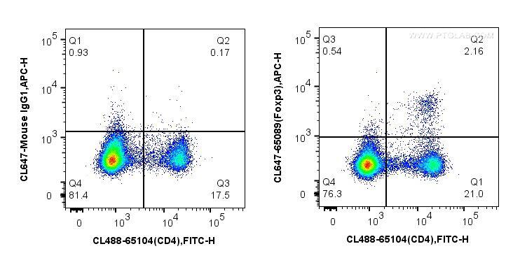
Flow Cytometry Perm Buffer 10x Pf00011 C Proteintech

Gating Strategy For Flow Cytometry A Schematic Illustration Of The Download Scientific Diagram

What Is Flow Cytometry Technology Networks

Combiflow Flow Cytometry Based Identification And Characterization Of Genetically And Functionally Distinct Aml Subclones Star Protocols
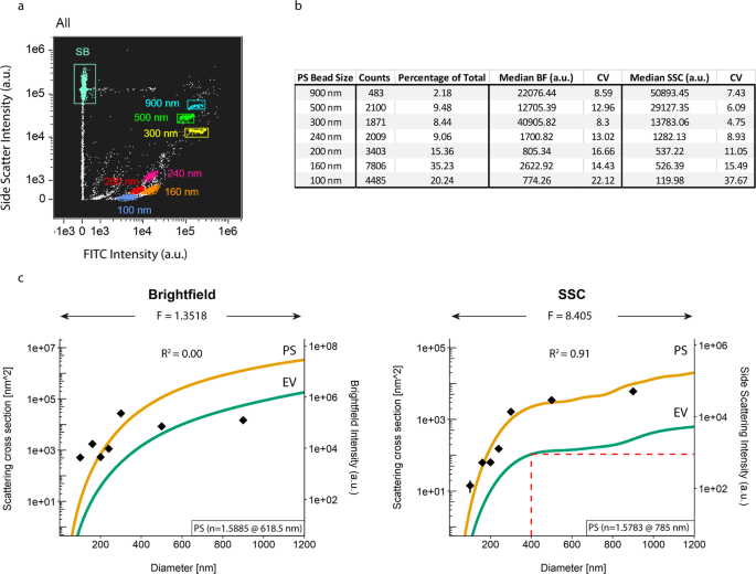
An Imaging Flow Cytometry Based Methodology For The Analysis Of Single Extracellular Vesicles In Unprocessed Human Plasma Communications Biology

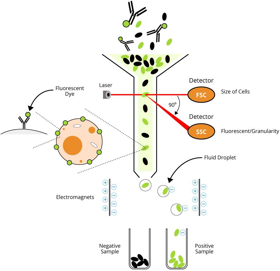
Flow Cytometry And Cell Sorting By Facs In The Flow Cell 1 The Download Scientific Diagram

Flow Cytometry Protocol For Staining Intracellular Molecules Using Detergents To Permeabilize The Cell Membrane R D Systems

Flow Cytometry Protocol For Staining Intracellular Molecules Using Alcohol To Permeabilize The Cell Membrane R D Systems
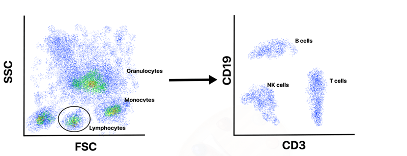
Analyzing Single Cells With Flow Cytometry

Representative Flow Cytometry Data From Experiments With Either Control Download Scientific Diagram

Flow Cytometry Based Protocols For Human Blood Marrow Immunophenotyping With Minimal Sample Perturbation Star Protocols

Flow Cytometry Facs Protocols Sino Biological
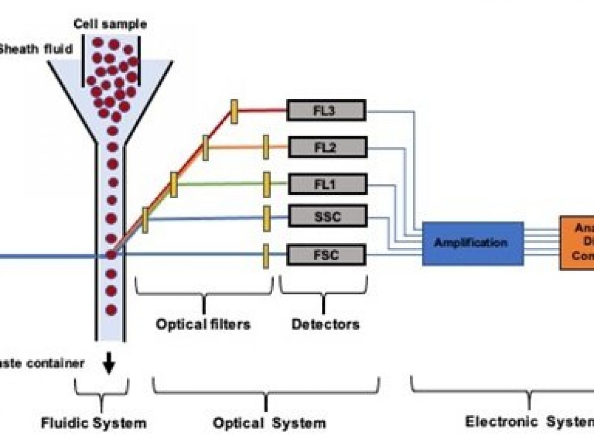
Cell Characterization Using Microfluidic Flow Cytometry Ufluidix
Flow Cytometry And Cell Sorting By Facs In The Flow Cell 1 The Download Scientific Diagram
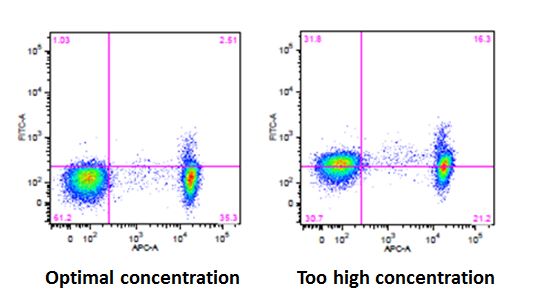
Blog 3 Considerations For Intracellular Flow Cytometry Icfc
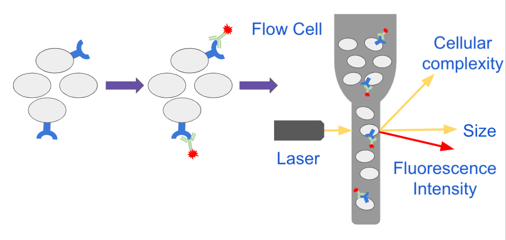
Analyzing Single Cells With Flow Cytometry

Gating Strategy For Flow Cytometry A Schematic Illustration Of The Download Scientific Diagram
